Research
This was the main project of my master's research at Zhejiang University. My work has focused on removing the key barriers that prevent multimode fibers (MMF) from becoming practical endoscopes. First, robustness under practical movements. Second, imaging speed. And finally, system integration toward in vivo clinical endoscopy.
Please find my selected publications and results below.
The long story starts here:
1. Introducation to miniaturized endoscopes
Endoscopy gives us optical access to regions that conventional microscopes cannot reach. But in many applications, the size of the imaging probe itself becomes the limiting factor.
In clinical endoscopy, conventional electronic gastroscopes are typically 5–10 millimeters in diameter. Imagine undergoing an endoscopic examination or surgery: ideally, the procedure should cause as little pain and discomfort as possible. In practice, however, conventional instruments can still cause considerable discomfort and often require local anesthesia or sedation. For this reason, manufacturers continue to develop thinner and more flexible devices. Some ultrathin endoscopes are now only around 3 millimeters in diameter, helping to reduce patient discomfort and tissue trauma. These endoscopes can provide real-time, wide-field image guidance and support flexible, multifunctional procedures. However, their resolution is insufficient to resolve cellular features, and they cannot image beneath the tissue surface. Another clinical approach is fiber-bundle-based confocal endomicroscopy. It reduces the imaging-probe diameter to approximately one to two millimeter while providing cellular-resolution images. However, the images suffer from honeycomb artifacts, the working distance is limited, and the probes are expensive and may need to be replaced after only 10–20 procedures.
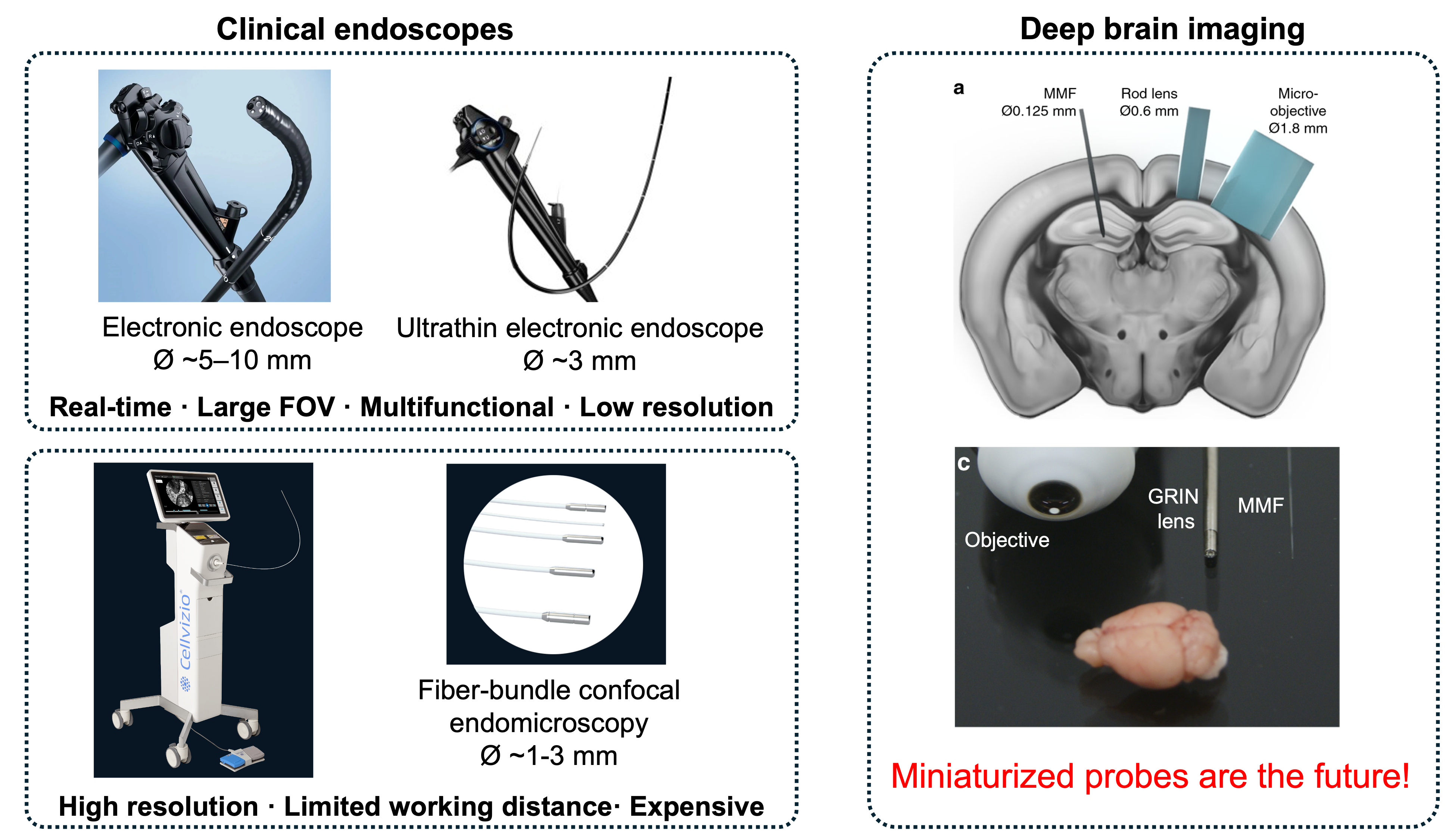
The size constraint becomes even more critical in neuroscience. Conventional microscope objectives can provide high-resolution imaging near the brain surface, but they are bulky and have a limited working distance. More importantly, brain tissue strongly scatters light, preventing an external objective from directly accessing deep brain regions with high spatial resolution. To overcome this depth limitation, GRIN-lens microendoscopesare inserted directly into the brain to relay images from deep structures back to the external microscope. This approach has enabled remarkable cellular-resolution imaging in freely behaving animals. However, GRIN lenses are typically around 0.5 to 1 millimeter in diameter, and inserting a probe of this size still displaces a substantial amount of brain tissue. It may also damage blood vessels, trigger inflammation, and disturb the neural circuits being observed. This motivates an interesting question: can we preserve high spatial information capacity while shrinking the probe by another order of magnitude? A multimode fiber offers exactly this possibility, potentially providing deep-brain optical access with substantially reduced tissue damage. Therefore, miniaturized probes are the future of endoscopic imaging.
2. Fiber imaging
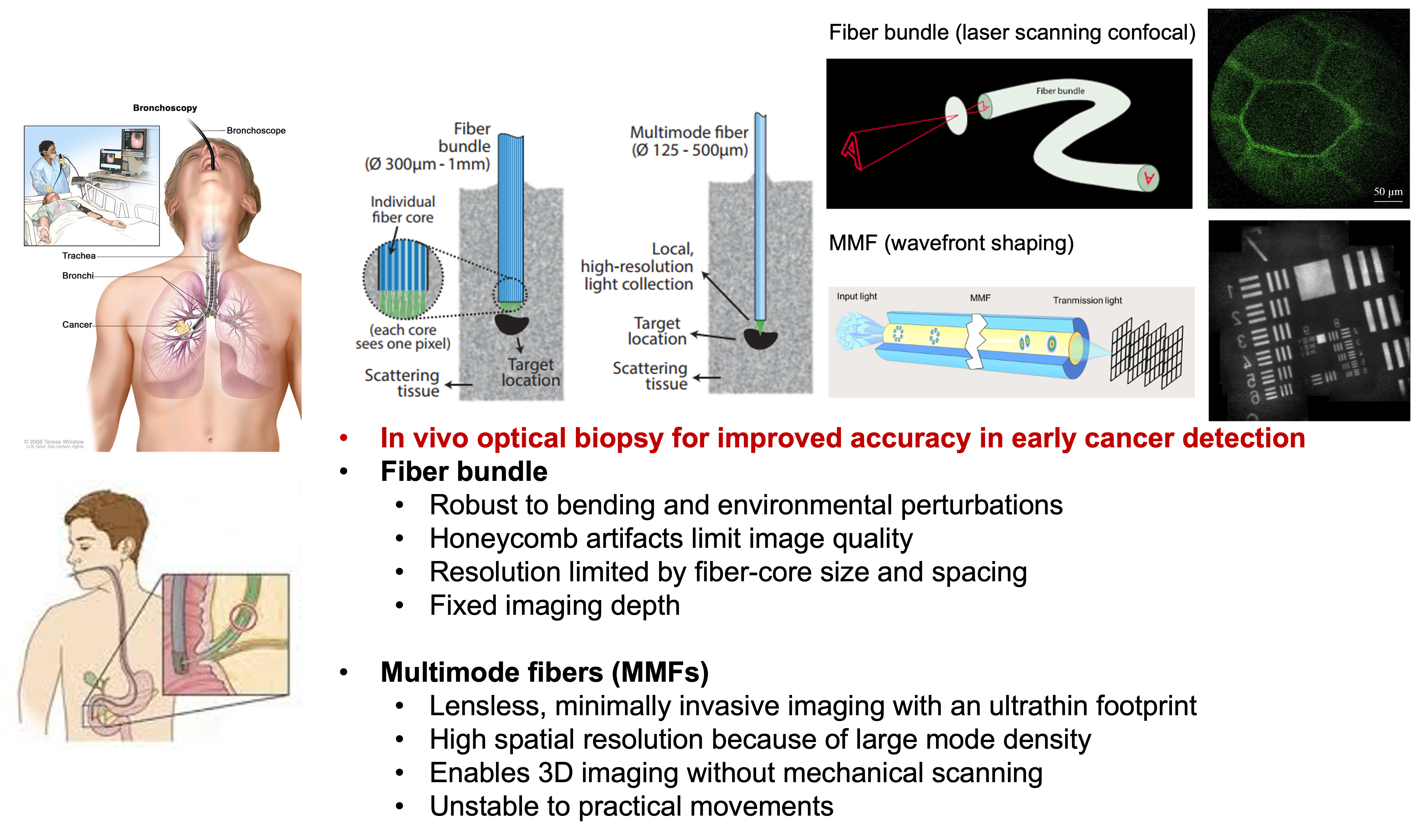
Conventional endoscopy often requires a tissue biopsy to be removed and sent to a laboratory for pathological analysis. This process takes time, delays diagnosis, and may require the patient to undergo an additional endoscopic procedure. Fiber imaging enables in vivo optical biopsy by providing high-resolution cellular and subcellular images directly inside the body, potentially supporting immediate diagnosis and more accurate early cancer detection. There are two main approaches to fiber-based imaging: fiber bundles and multimode fibers (MMF). Fiber bundles directly transmit images through thousands of individual cores and are relatively robust to bending. However, they suffer from honeycomb artifacts, fiber-core spacing limits their spatial resolution, and their short, fixed working distance restricts the accessible imaging depth. In contrast, multimode fibers enable lensless, minimally invasive imaging through an ultrathin probe approximately 100 μm in diameter. Their large mode density supports high spatial resolution, while wavefront shaping enables 3D imaging without mechanical scanning. However, their performance is highly sensitive to real-world fiber movement and deformation.
3. STABLE
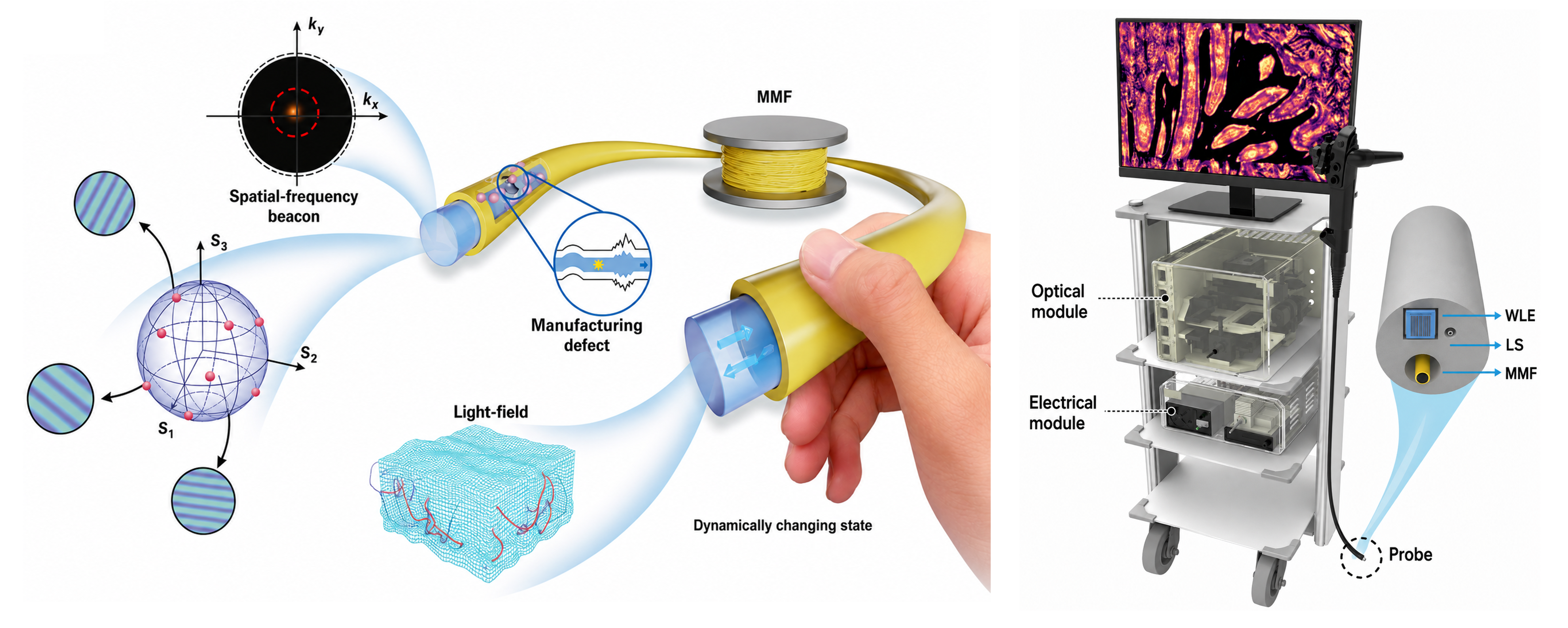
In our Nature Photonics paper, we proposed a method named 'STABLE' (Spatial-frequency tracking adaptive beacon light-field-encoded endoscopy) that enables stable MMF imaging under practical movements for the first time. There are four key contributions of this paper:
- Full-vector modulation (FVM):
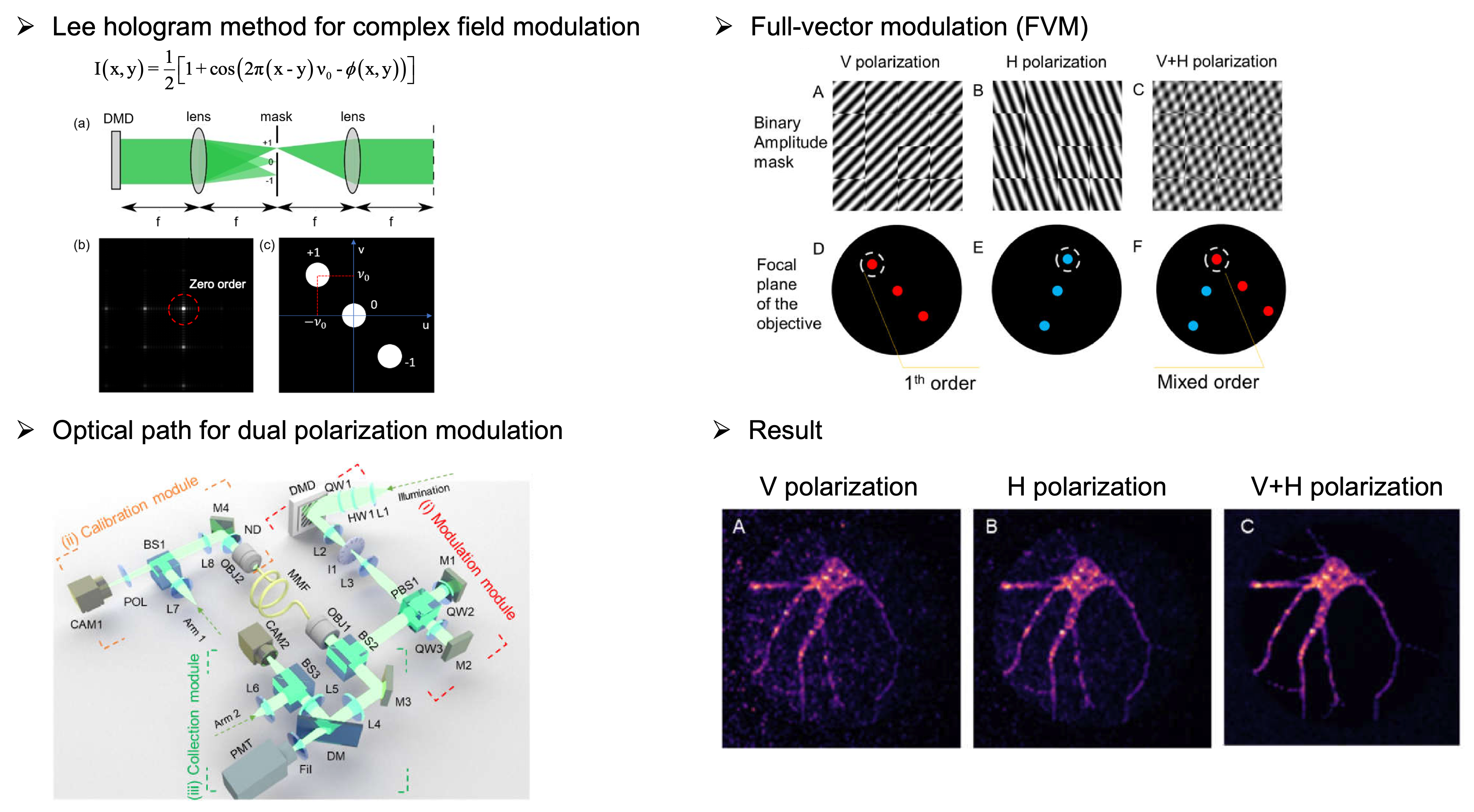
Strong mode and polarization coupling occurs in long and bent MMFs. Therefore, full-vector modulation is crucial for effectively controlling light propagation through the fiber. We developed a method that uses a single binary DMD to simultaneously modulate the amplitude, phase, and polarization of the optical field at the MMF input facet.
Full-vector modulation with a single binary DMD. - Reflection beacon for fast TM correction:
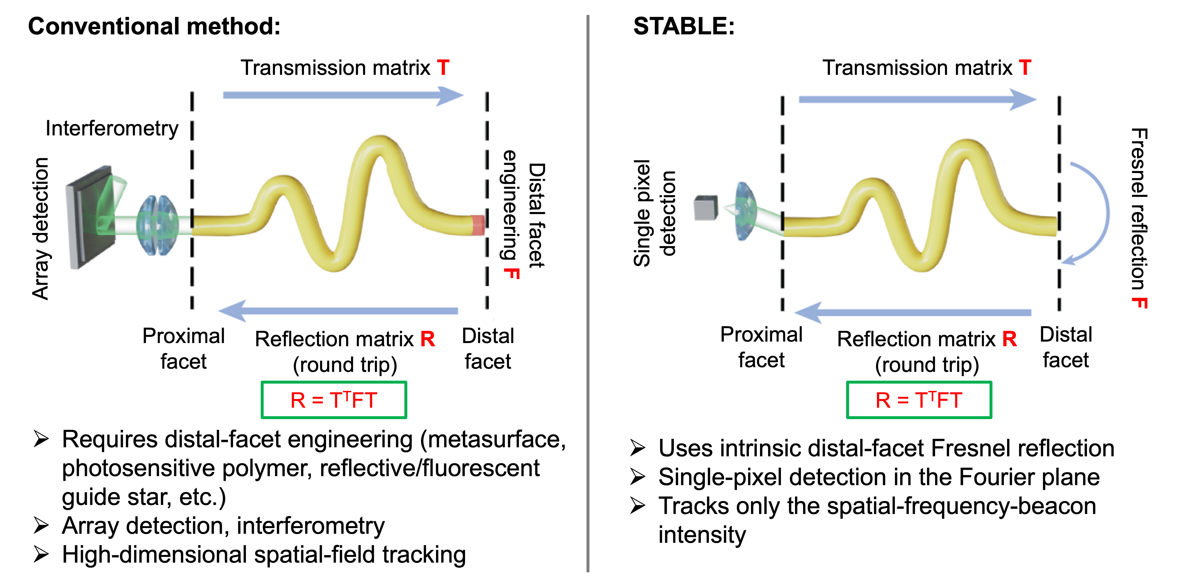
The transmission matrix (TM) of a MMF depends strongly on its shape. Once the fiber bends or moves, the original calibration is no longer valid. For practical endoscopic imaging, we therefore need an efficient way to identify the current TM using measurements from only the proximal side, without accessing or modifying the distal facet.
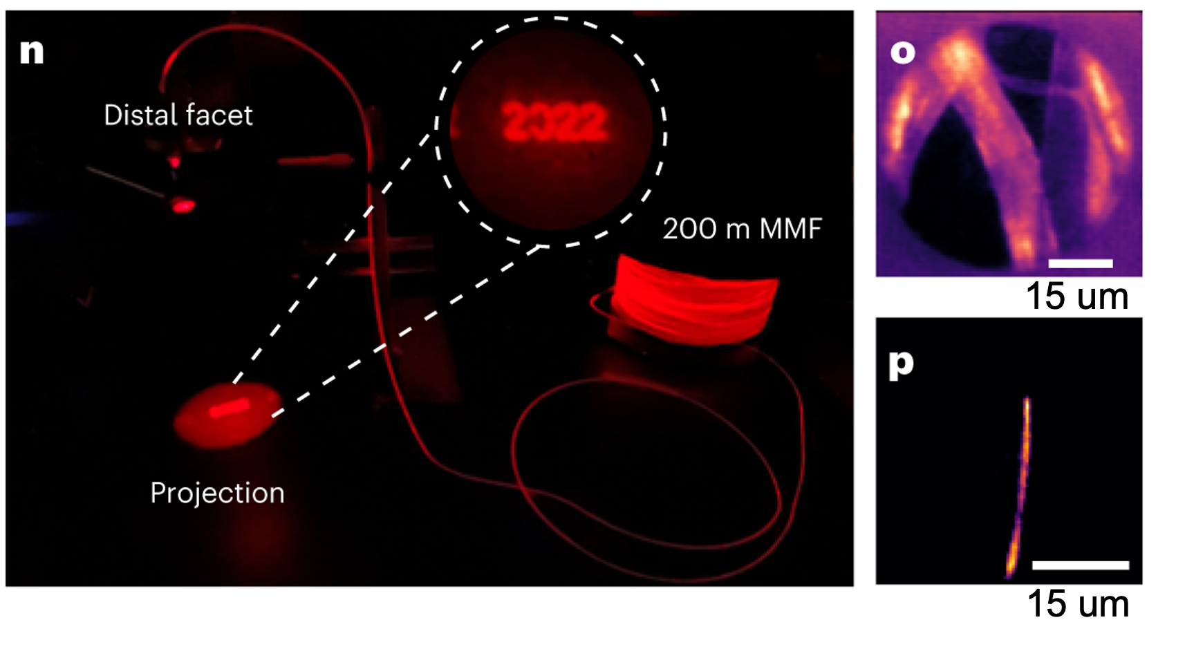
Our STABLE method uses the Fresnel reflection from the distal fiber facet as an intrinsic guide signal. During pre-calibration, we measure the single-pass TM and the corresponding round-trip reflection matrix (RM) for several representative fiber configurations, creating a library of paired TMs and RMs. During operation, an RM-derived wavefront propagates through the fiber, undergoes Fresnel reflection at the distal facet, and travels back through the fiber. The returned field is then mapped into the spatial-frequency domain at the proximal side. When the tested RM matches the current fiber state, the reflected light is concentrated into a sharp frequency-domain focus, which serves as the reflection beacon. We identify the RM that produces the highest beacon intensity and select its paired TM for imaging.
In this way, STABLE converts high-dimensional optical-field tracking into simple single-pixel beacon-intensity tracking. It requires neither distal access nor an engineered reflector, while improving tracking speed and sensitivity. Moreover, the calibration library can remain compact because MMFs exhibit memory effects and translational and rotational degeneracies: different fiber configurations can produce very similar TMs. In our experiments, approximately 20 TM–RM pairs were sufficient to cover the tested fiber states.
STABLE imaging via a spatial-frequency reflection beacon and a TM-RM library. - 3D & super-resolution imaging with bend MMF:
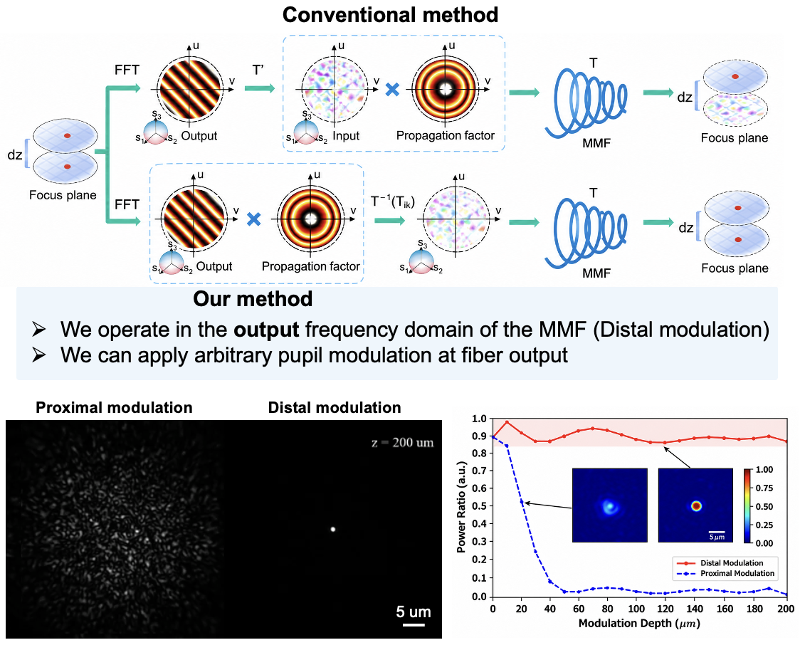
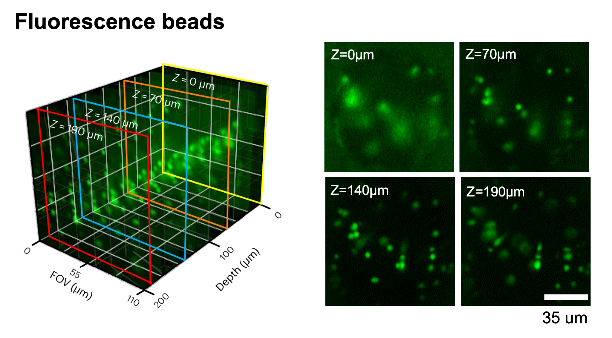
Traditional 3D digital refocusing shifts the focal plane by applying a quadratic phase in the input spatial-frequency domain. However, in a long or bent multimode fiber, strong mode coupling mixes the angular spectrum. As a result, the angular spectrum is no longer propagation-invariant, and the transmission matrix is not diagonal in the spatial-frequency basis. The focus therefore degrades significantly as the imaging depth increases. To overcome this limitation, we define the desired optical field directly in the output spatial-frequency domain of the MMF and calculate the corresponding input wavefront through phase conjugation. This approach maintains nearly unchanged focus quality up to 200 μm from the distal fiber facet. It also enables arbitrary pupil/wavefront modulation at the fiber output.
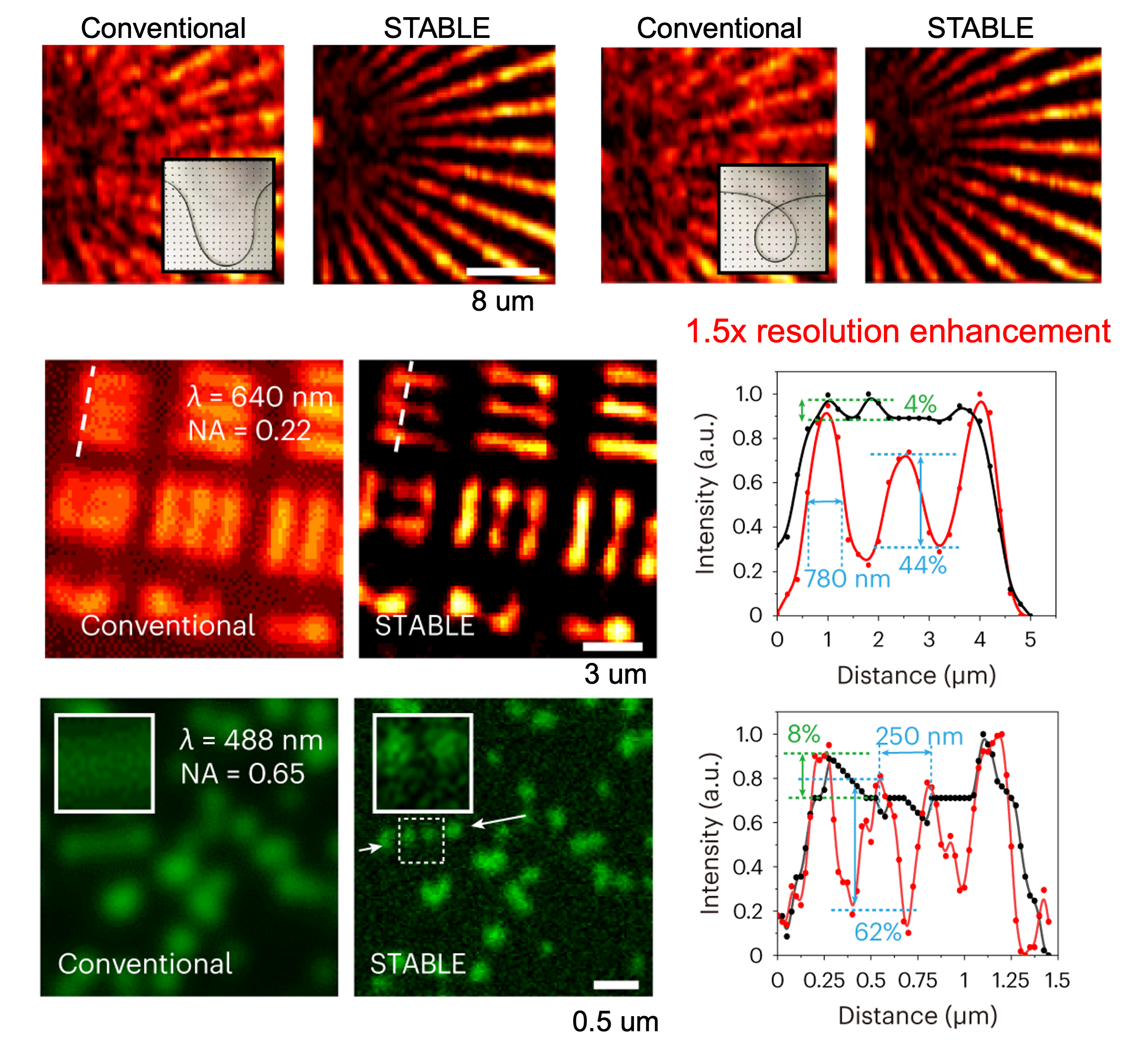
3D focusing operating from fiber output frequency domain. We further applied fluorescence-emission-difference (FED) imaging to achieve a 1.5-fold improvement in spatial resolution. The super-resolved image was reconstructed by subtracting the doughnut-beam-scanned image from the Gaussian-beam-scanned image, with the doughnut beam generated by applying a vortex phase at the MMF output pupil.
Super-resolution imaging with MMF. - Practical applications: clinical integrated endoscopy; living mice
We integrated an MMF imaging probe into an ultrathin electronic endoscope to demonstrate a compact multimodal endoscopic platform. The electronic endoscope provides wide-field guidance for navigation through the narrow airways of a bronchial model, while the MMF probe provides autofocusing and high-resolution imaging of fine tissue features.
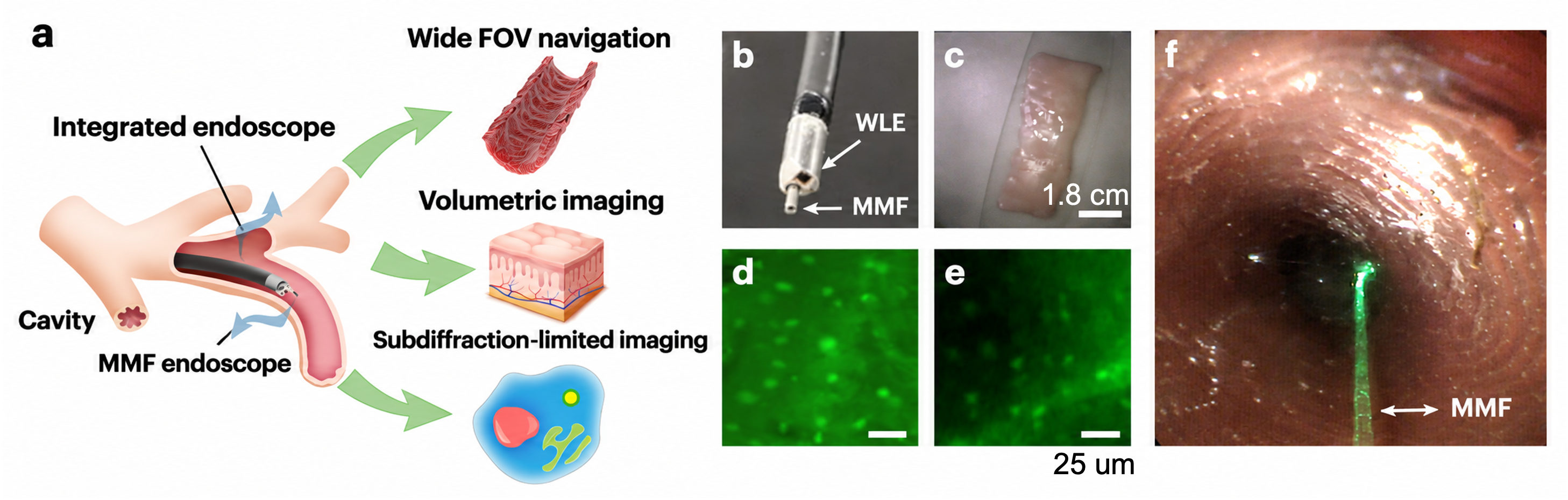
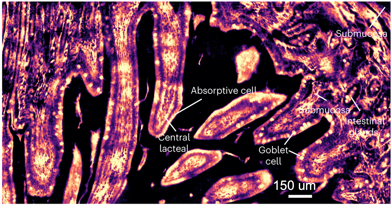
Integrated electronic and MMF endoscopy. Finally, we demonstrated in vivo imaging of the mouse gastrointestinal tract using the integrated endoscope across multiple GI organs. STABLE enabled subcellular-resolution imaging of the mucosa, as well as subsurface tissue imaging through direct insertion of the MMF probe. By positioning the probe approximately 400 μm beneath the tissue surface, we observed clear structural differences between healthy and cancerous colon tissue.
Image Gallery
4. 3D compressive imaging through MMF
The imaging speed of MMF-based systems is fundamentally constrained by the point-by-point scanning scheme, the modulation speed of spatial light modulators, and the Nyquist sampling requirement. For example, for an MMF with a 50-µm diameter and an NA of 0.22, diffraction-limited imaging requires a lateral scanning interval of approximately 0.6 µm, corresponding to at least 84 × 84 sampling points across the full MMF field of view. Even with a high-speed DMD, this limits the maximum imaging rate to approximately 3.2 Hz for a single 2D plane. This limitation becomes substantially more severe for 3D imaging, where conventional approaches require sequential layer-by-layer scanning.
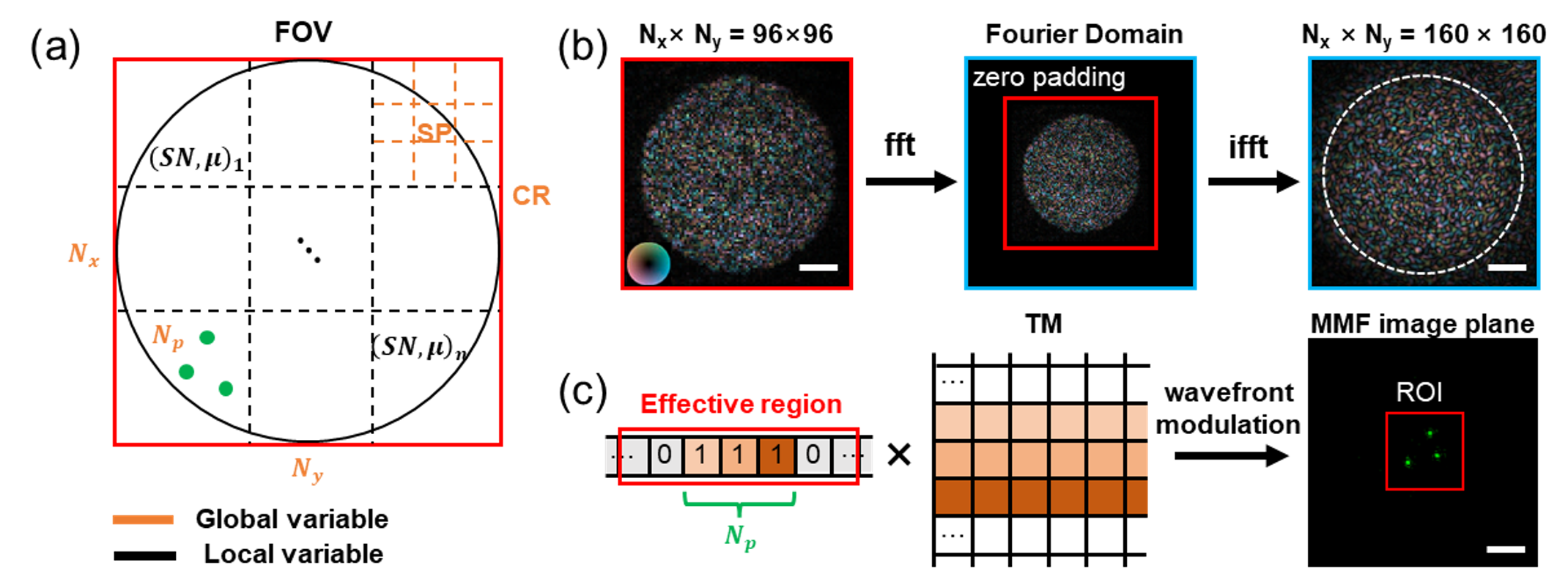
To overcome this limitation, we introduced compressive sensing into the scanning framework of MMF imaging. We designed a more efficient 3D scanning basis consisting of multiple focal spots distributed throughout the imaging volume, and developed a computational reconstruction algorithm to solve the corresponding 3D inverse problem. There are three key contributions of this work:
- We demonstrated that a focal-spot basis outperforms a speckle basis due to its improved orthogonality. We further found that using multiple focal spots in each illumination pattern leads to better reconstruction performance.
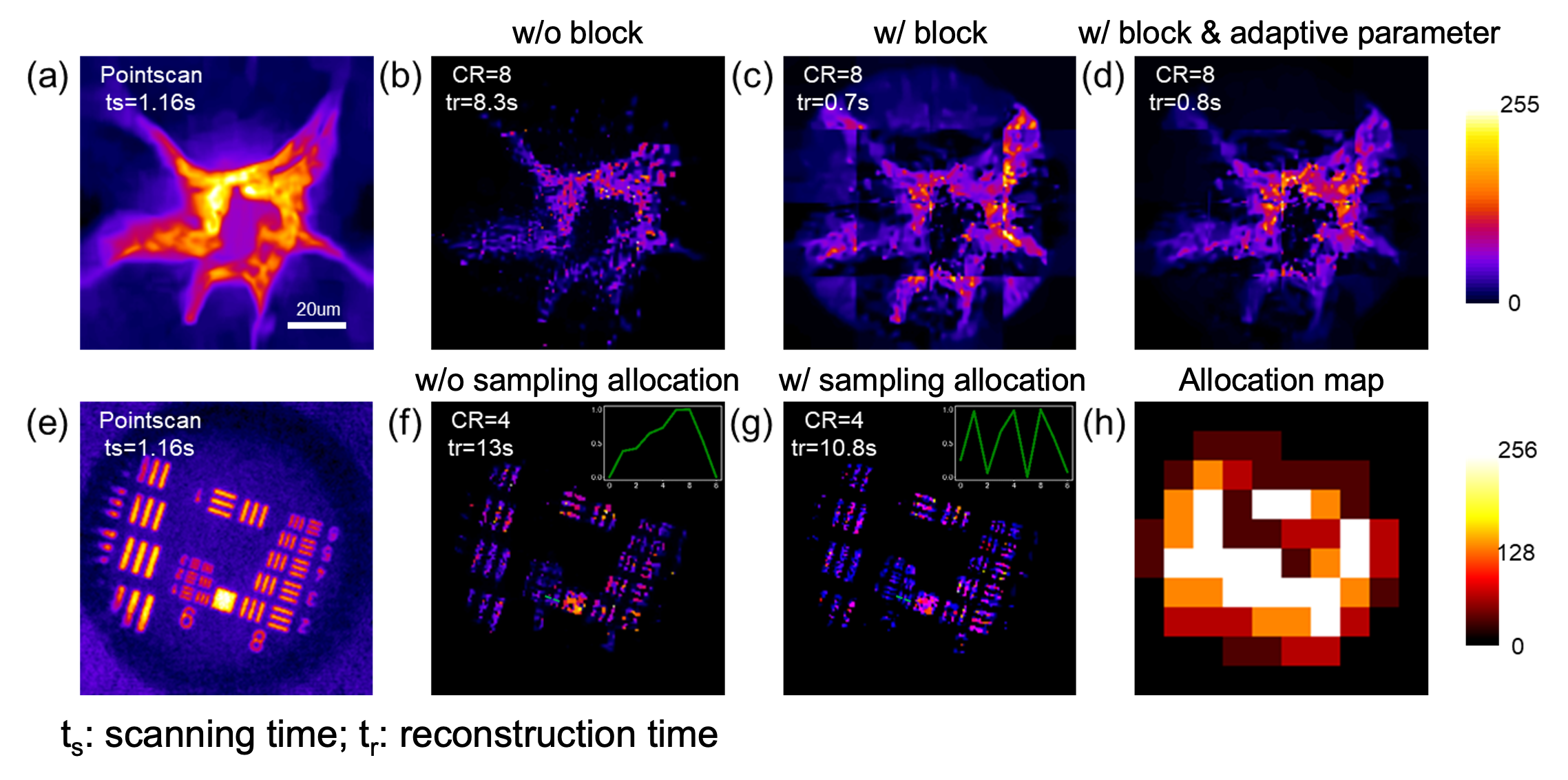
- We developed a block-wise scanning scheme with adaptive parameter adjustment for each block, enabling parallel reconstruction and efficient allocation of sampling measurements across different regions. Experimentally, we demonstrated an eight-fold improvement in acquisition speed on lung tissue sections and fluorescent bead samples.
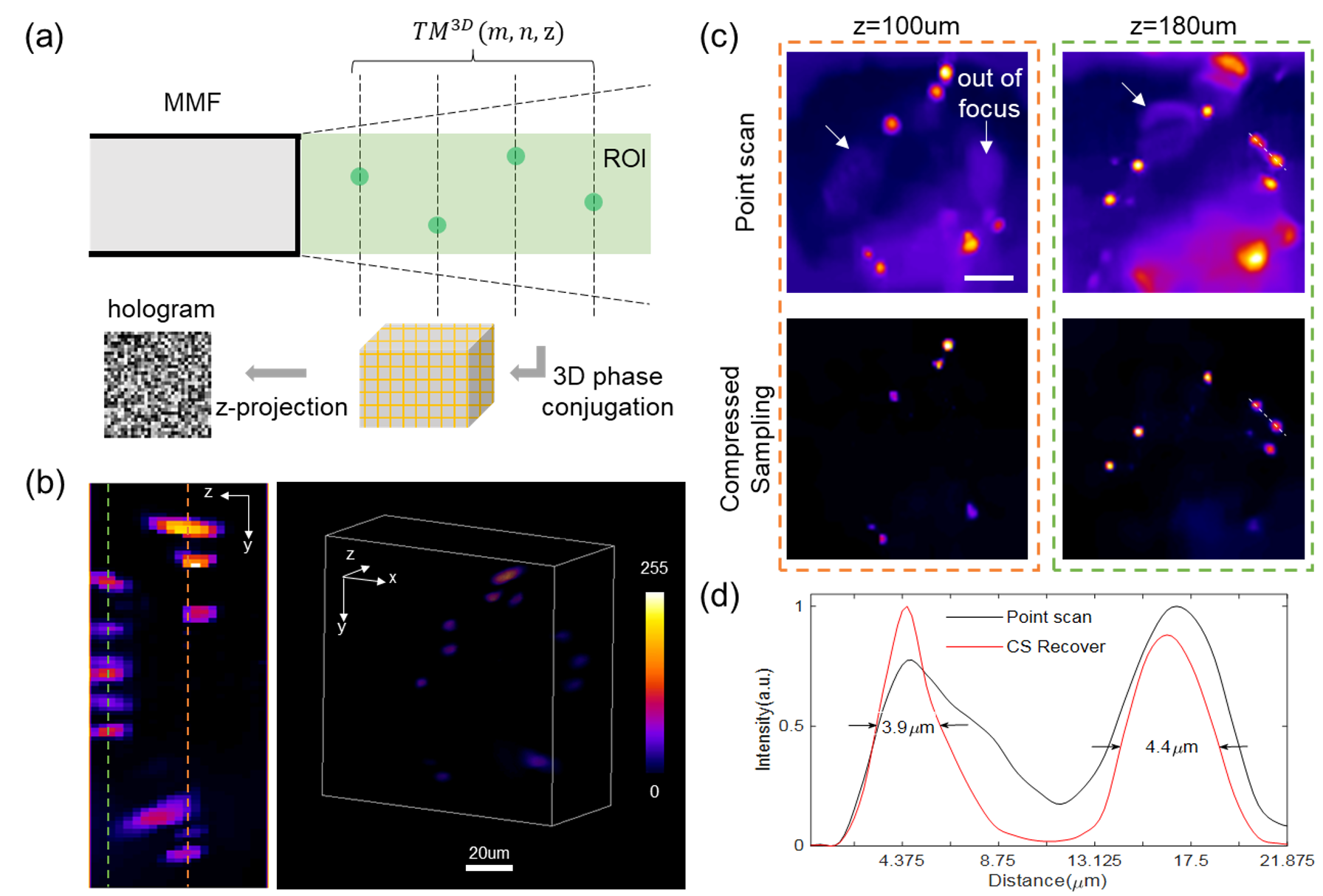
- We designed a multiple-focal-spot sampling basis directly in 3D, rather than relying on conventional layer-by-layer scanning. By solving the corresponding 3D inverse problem, this approach enables computational 3D deconvolution and suppression of out-of-focus signals. We achieved 3D computational imaging over a 100 × 100 × 200 μm³ volume with an acquisition time of only 1.7 s, representing a nine-fold improvement in imaging speed compared with conventional point-by-point scanning.
Image Gallery
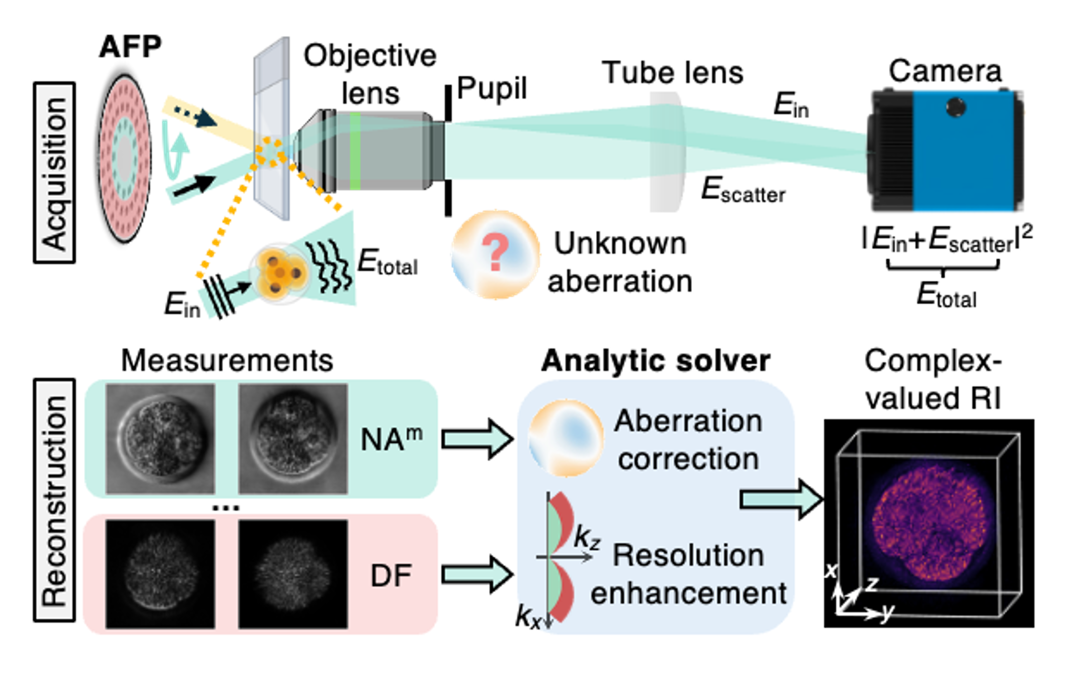
This is the main project of my PhD research. I focus on label-free refractive index (RI) tomography of thick 3D specimens. RI tomography enables quantitative 3D visualization of biological structures based on their intrinsic optical properties, without the need for fluorescent labeling. In particular, I develop non-interferometric RI tomography techniques that rely on intensity-only measurements, providing a simple, robust, and phase-stable approach well suited for long-term imaging of living biological samples.
- Computational imaging: I developed computational imaging techniques to address optical aberrations, improve 3D resolution, and mitigate multiple-scattering effects, thereby enhancing both data acquisition efficiency and reconstruction quality.
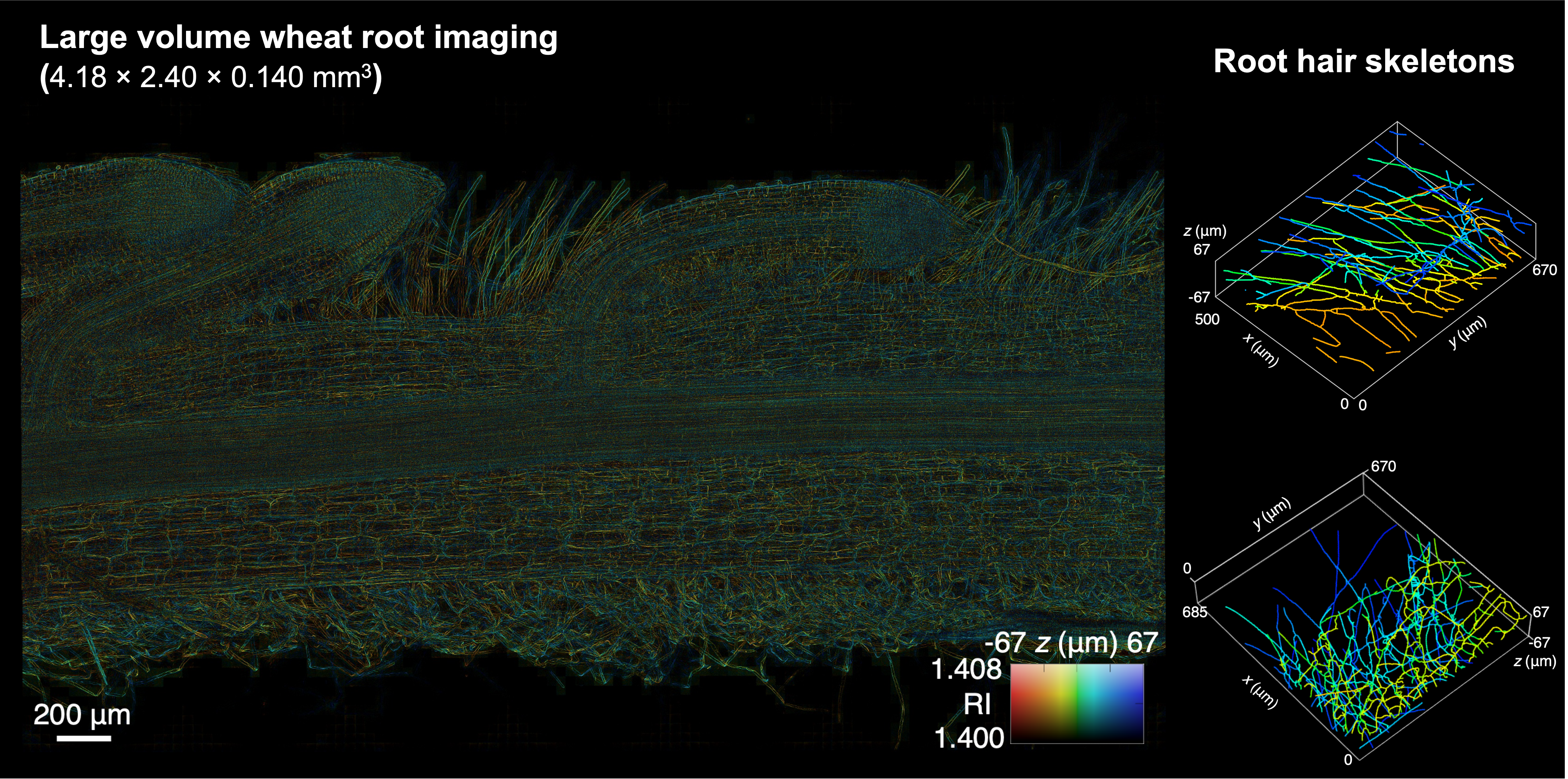
- Biomedical imaging: I apply these techniques to high-resolution, label-free 3D imaging of biological specimens across a broad range of applications, including mouse oocytes, mouse and human preimplantation embryos, mouse brain tissue, plant roots, pathology slides, and algae.
- Clinical translation: I am currently collaborating with a start-up company to develop a device called LANTERN, which aims to provide high-quality, label-free, time-lapse imaging of human embryos in in vitro fertilization (IVF) clinics. We hope this technology can provide valuable insights into early embryonic development and assist embryologists in assessing embryo health and developmental potential.
Please find my related publications and selected results below.
Image Gallery
I worked on this as a side project during the first two years of my PhD research.
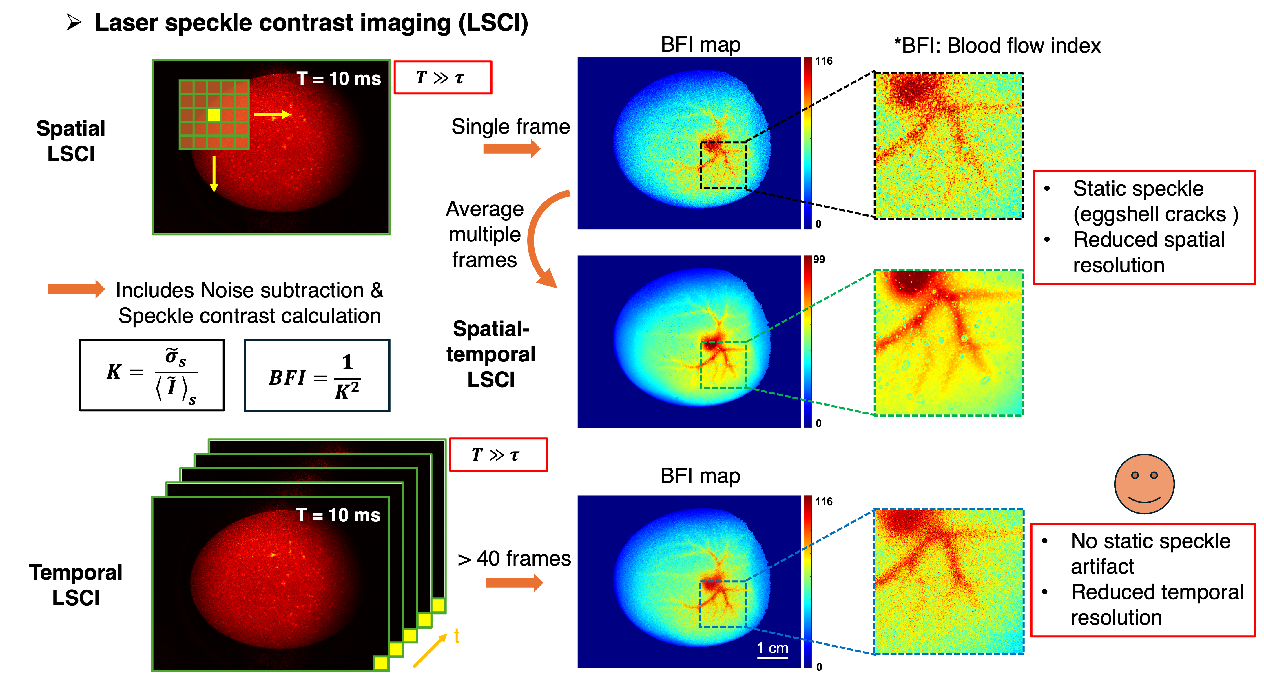
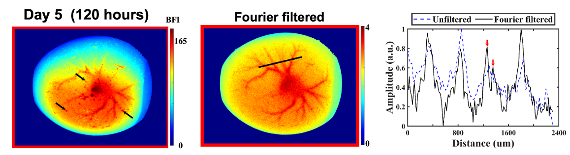
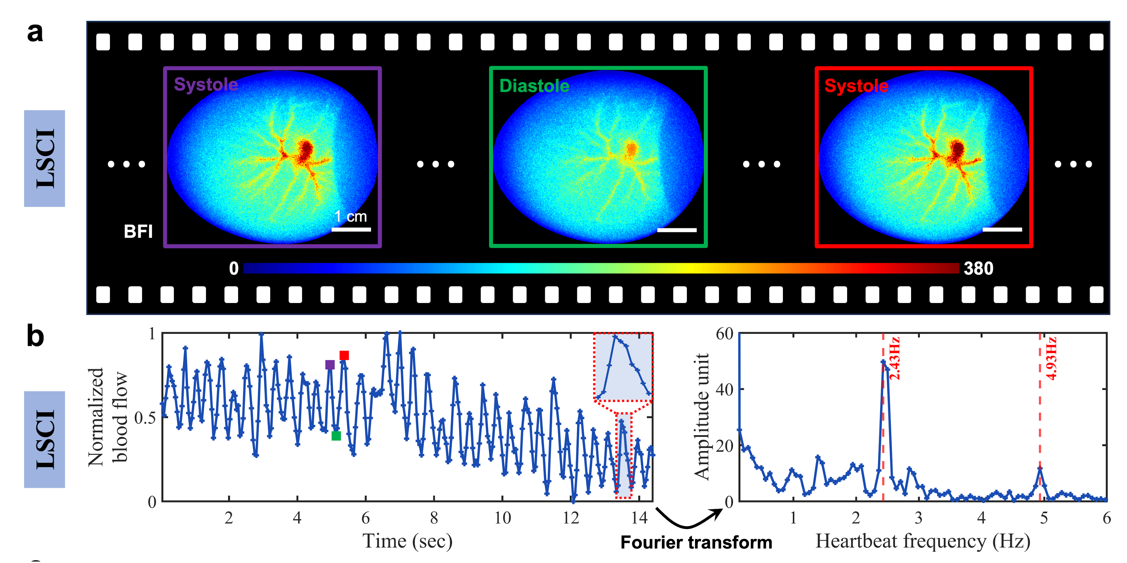
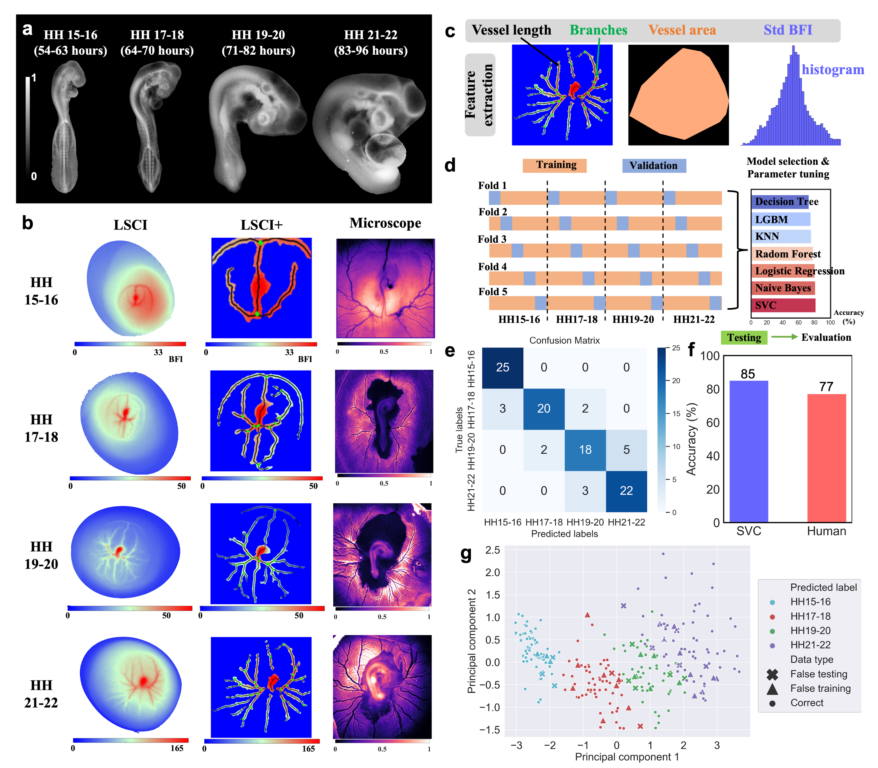
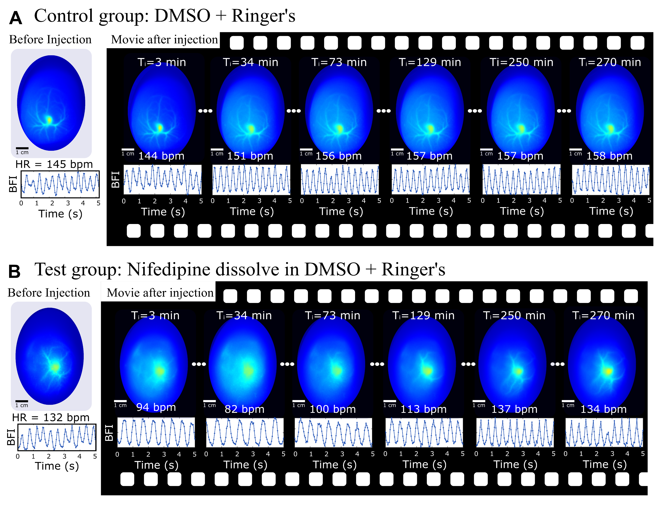
Imaging blood vessels in early-stage avian embryos has a wide range of practical applications, including developmental biology studies, drug and vaccine testing, and early sex determination in agriculture. Optical imaging methods, such as brightfield transmission imaging, offer a compelling solution due to their use of safe, non-ionizing radiation and operational simplicity. However, imaging through intact eggs remains challenging due to eggshell opacity and strong light scattering.
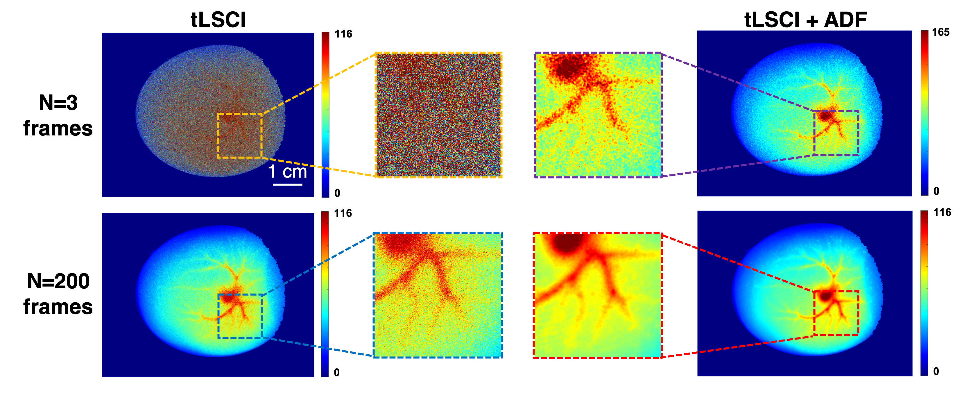
In this project, I revisited and modified laser speckle contrast imaging (LSCI) to enable high-quality, artifact-free, real-time visualization of blood-flow dynamics non-invasively through intact eggshells.
Selected publication as follows:
Image Gallery

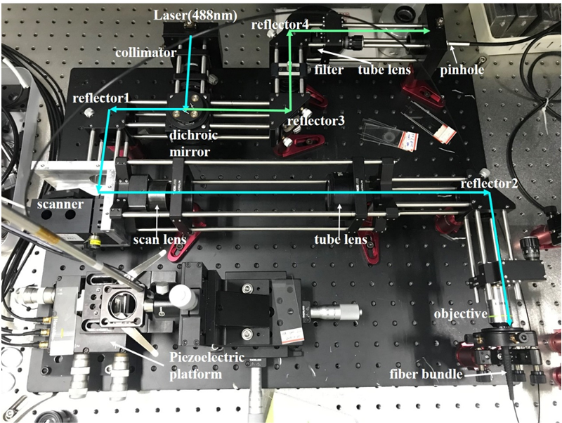
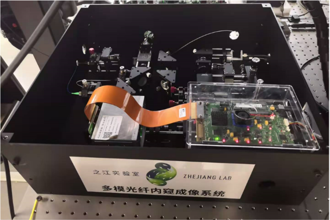
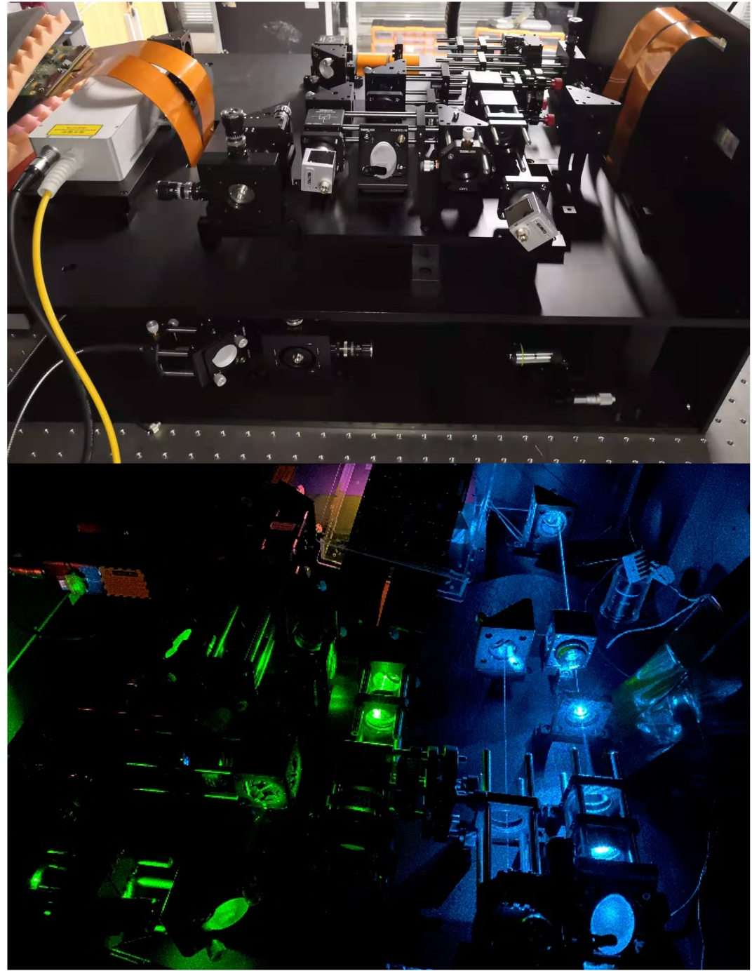
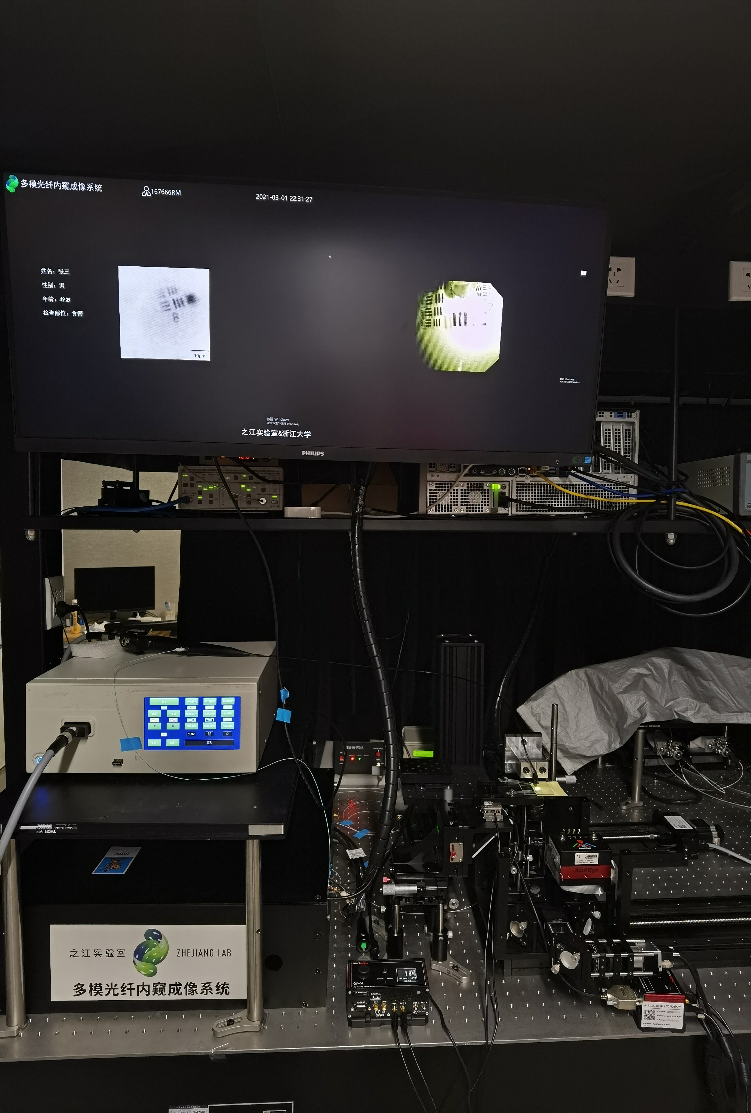
This section showcases a collection of optical imaging systems that I have designed and built.
Image Gallery